
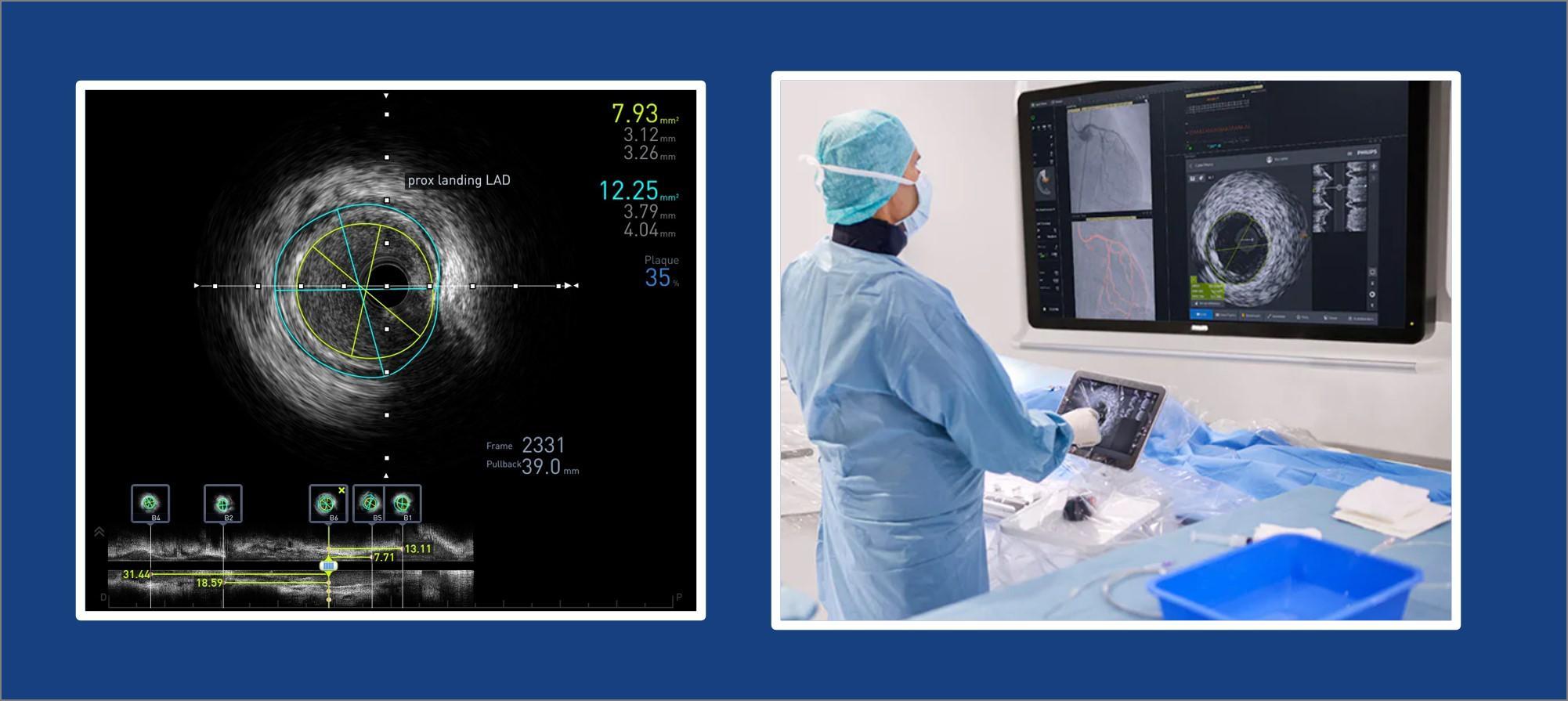
What is Intravascular Ultrasound (IVUS)
Intravascular Ultrasound (IVUS) allows us to see a coronary artery from the inside-out. When used to evaluate the coronary arteries, IVUS can show the entire artery wall and provide important information about the amount and type of plaque buildup, which can help determine if you are at risk for heart attack.
Ultrasound does not use ionizing radiation, has no known harmful effects, and can provide clear pictures of soft tissues that are not well seen on x-ray images.
Cardiologists feel that the new information yielded by IVUS can make a significant difference in how a patient needs to be treated, information about accurate stent placement and thereby reducing complications.
How does IVUS work?
IVUS uses high-frequency sound (called ultra sound) waves to provide images from inside the blood vessels. Actually IVUS uses echocardiography: the same technology as the ultrasound imaging used in treadmill tests and many other medical exams.
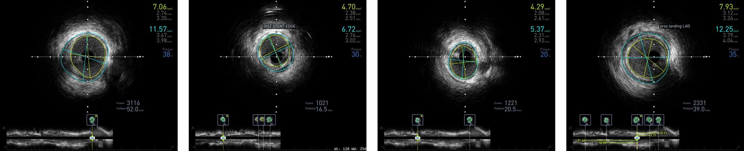
The transducer on the end of the catheter uses sound waves to produce pictures of the blood vessels. Doctors can move the catheter to obtain images of the inside of the vessels at different locations. It becomes, in effect, a tiny camera that gives us a cross-sectional view of the artery, a view that shows distinct circular layers, using shades of gray or colors.
How is IVUS Different from Standard Angiography?
Although angiography shows “narrowings”, as well as a dynamic picture of the blood flow, it does not differentiate the other layers. In several situations, the increased information provided by Intravascular Ultrasound literally can change the picture of the disease, and affect treatment decisions.
Sometimes the plaque pushes deeper into the vessel wall, giving the appearance that the lumen is not significantly blocked. Yet a significant amount of diseased plaque may exist within the arterial wall, ready to rupture and cause a cascade of events, resulting in a heart attack. This is called vulnerable plaque, and cannot be visualized using standard angiography.
With the accurate measurements of both the true diameter of the artery and the diameter of the open lumen channel provided by IVUS, the guesswork is taken out of choosing the correct size balloon and stent. Using only angiography, a cardiologist may underestimate the size of a diseased artery.
IVUS has many benefits including:
showing the presence and amount of plaque in arteries
providing information about what the plaque is made of
more accurate stent placement
no exposure to ionizing radiation
finding areas of vein external compression
finding stenosis or narrowing which is not well seen with angiography